Established 1996 — Advancing Ultrasound Education in Healthcare
The Hameed Gul Memorial Ultrasound Institute was founded in 1996 with the vision of providing high-quality diagnostic ultrasound education to a wide spectrum of healthcare professionals. The institute has played a pioneering role in promoting ultrasound awareness and practical training across diverse healthcare roles including:
Our Impact
With nearly three decades of excellence in medical ultrasound training, the Hamid Gul Memorial Ultrasound Institute continues to empower healthcare professionals with diagnostic skills that make a real difference in patient care. Our alumni are a testament to the global impact of our programs.
International Academic Collaborations
From its early years, the Hameed Gul Memorial Ultrasound Institute recognized the importance of international collaboration in advancing medical ultrasound education. The institute initially established an academic partnership with the Burwin Ultrasound Institute, Canada, which laid the foundation for a globally informed curriculum and introduced students to international best practices in diagnostic ultrasound.
Building on this momentum, the institute later developed a prestigious academic collaboration with the Jefferson Ultrasound Research and Educational Institute (JUREI), a world-renowned center for ultrasound education and research. At the time, JUREI was under the leadership of Prof. Dr. Barry B. Goldberg, a pioneer in the field of medical ultrasound. Prof. Goldberg was among the earliest radiologists to adopt and promote ultrasound technology globally. His career included work on one of the very first ultrasound machines, making him a historical figure in the evolution of modern diagnostic imaging.
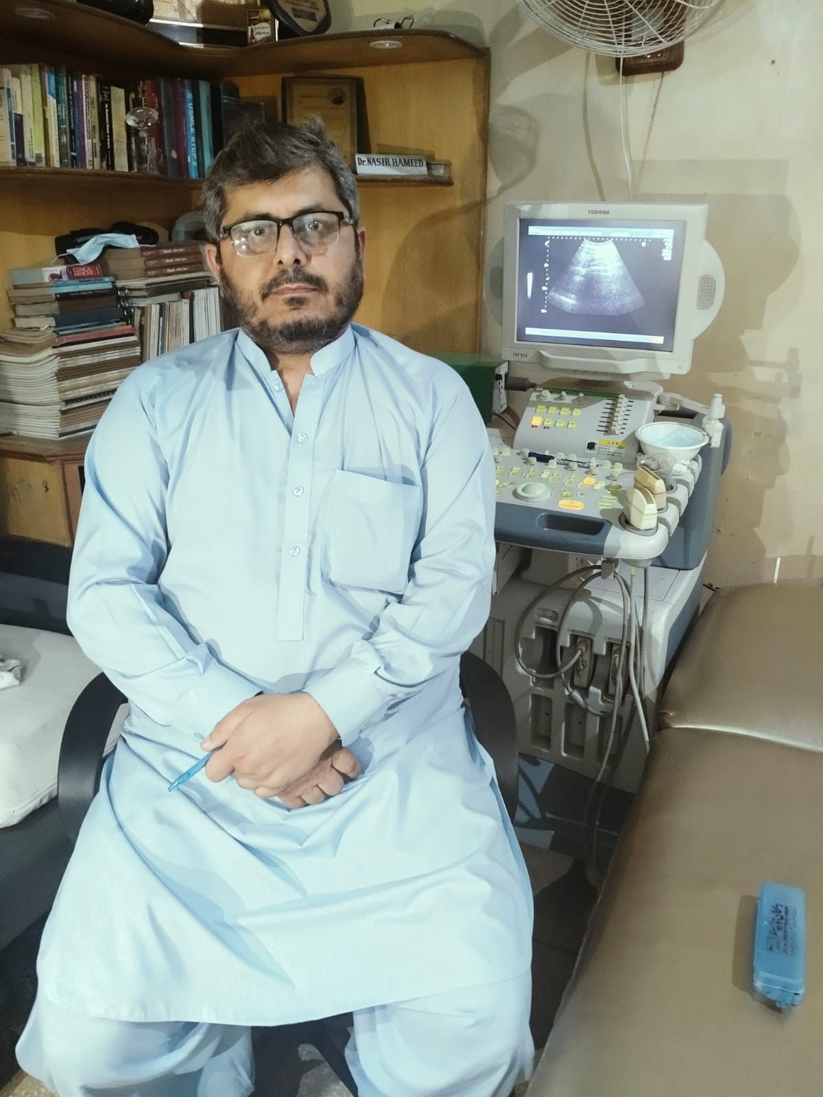
The founder of the Hameed Gul Memorial Ultrasound Institute, Dr. Nasir Hameed, had the distinct honor of meeting and working with Prof. Dr. Goldberg during his visiting fellowship at JUREI in 2003.
This partnership was facilitated through JUREI’s Middle East branch, which was headed by Prof. Dr. Syed Amir Gilani, an influential figure in the regional advancement of ultrasound education. Prof. Gilani later became Professor and Vice Dean at the University of Lahore, where he continued to champion ultrasound education and research. His academic journey eventually led him to the University of Sharjah in the United Arab Emirates, where he served as a Professor of Radiology.
These collaborations not only enhanced the academic stature of the institute but also ensured that students and faculty had access to cutting-edge research, global teaching methodologies, and exposure to international diagnostic protocols. The impact of these partnerships continues to be felt today in the caliber of training and the professional accomplishments of the institute’s alumni.
Our First Student and Early Success
The first student to enroll was Dr. Nausheen Fahim, who joined the Basic Ultrasound Course in 1997. Since then, the institute has trained over 500 medical professionals, most of whom are qualified doctors. Many were working as training medical officers at the time of enrollment, while others were senior consultants, including professors. Some of the names to mention here include:
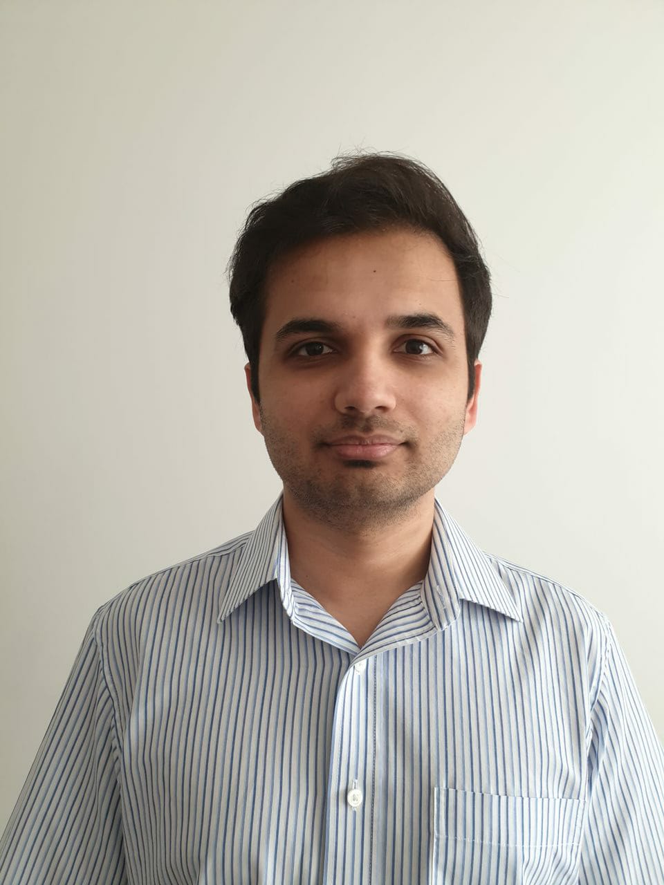
Our Faculty
Learn from industry-leading professionals with extensive experience in clinical practice and education.
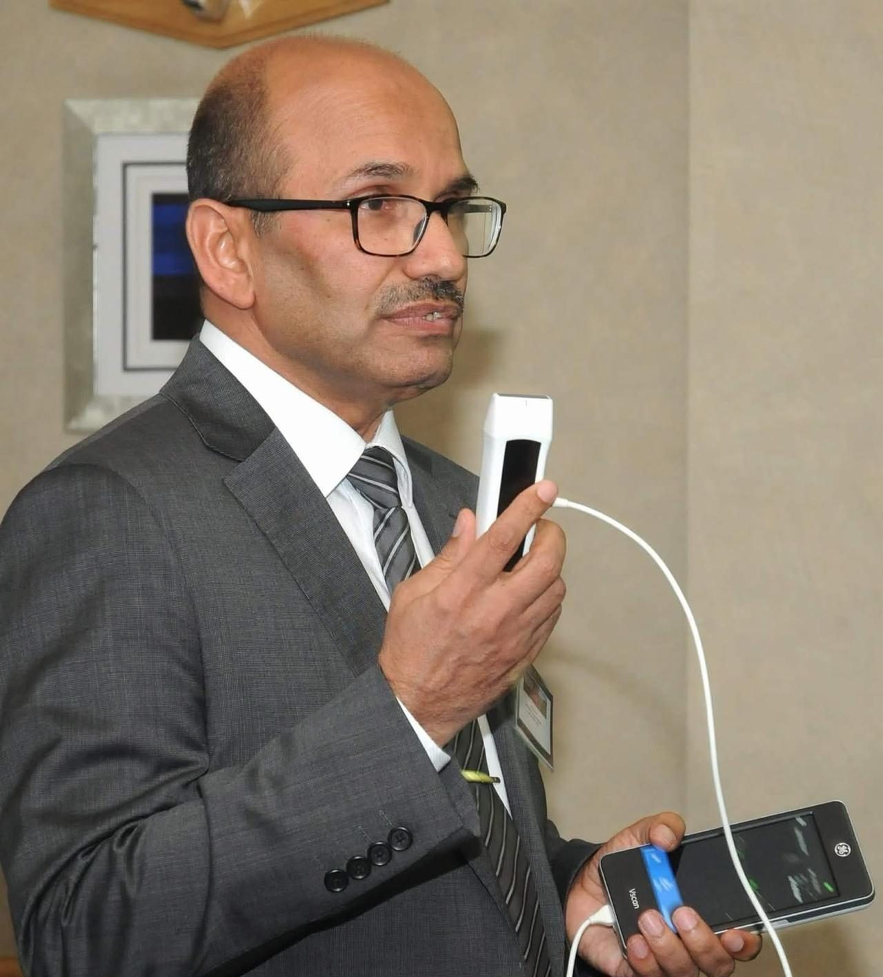
Medical Education Director
M.B.B.S (Pak) PhD Medical Ultrasound (UOL PAK)
PG Diploma in Medical Education (UK)
Associate Fellow, Higher Education Academy (UK)
Associate Professor in Medical Imaging UCLAN (UK)
International Faculty
Windsor House Group Practice Leeds, United kingdom
MBBS (Pakistan), Diploma in Medical Diagnostic Ultrasound, MRCGP (UK)
Abdominal & Small Parts Ultrasound Specialist
Senior Specialist (Medical Ultrasound)
M.B.B.S (Pak),
P.G Diploma Ultrasound (Pesh),
M.Phil Ultrasound (UOL Pak), ICEAF (USA)
Ultrasound specialist
M.B.B.S (Pak), P.G Diploma Ultrasound (Pesh)
Ultrasound specialist
FCPS(TMO)Paediatric, 2022 – Present, LRH Peshawar
Lecturer in Biochemistry 2021-2022-Khyber Medical College
MO in Surgical ICU 2020-2021-Northwest medical Hospital
MBBS— Peshawar Medical college (2019)
MD
Basic Ultrasound Course 2008
Ultrasound Diploma (Pak)
Ultrasound Scanning Experience: 17 Years

Ultrasound Specialist/Practical Ultrasound Supervisor
MBBS (Pesh)
RMP (PMDC)
Ultrasound Diploma (Pak)
International Faculty
Consultant Radiologist
Tameside and Glossop Integrated Care NHS Foundation Trust
Ashton-under-Lyne, OL6 9RW
United Kingdom
- FRCR – Clinical Radiology, Royal College of Radiologists, London, UK (2002)
- CCST – Clinical Radiology (2004)
- FCPS – Diagnostic Radiology, College of Physicians & Surgeons, Pakistan (1997)
- MCPS – Diagnostic Radiology, College of Physicians & Surgeons, Pakistan (1995)
- MBBS – Khyber Medical College, University of Peshawar, Pakistan (1986)
Professor
University of Sharjah
Sharjah, Emirate, UAE
- Pro – Reactor Health & Allied Sciences Lahore
- Professor & Dean Faculty Health & Allied Sciences (University of Lahore)
- Associate Professor Allied & Health Sciences (Alzaiem Alazhari, university, khartoum, Sudan)
- Head of Dep – AFRO – Asian Institute of Radiology Lahore
Courses
Master the Fundamentals of Ultrasound
Basic Ultrasound Course
Offered by Hameed Gul Memorial Ultrasound Institute
Eligibility: Medical Doctors with MBBS or MD Qualification
Take the first step towards a skilled and confident practice in diagnostic ultrasound. Our Basic Ultrasound Course is designed specifically for qualified medical doctors who are looking to gain core competencies in ultrasound imaging through a structured, hands-on program.
Program Highlights
Introduction to Ultrasound Physics
• Sound wave principles
• Image formation and resolution
• Knobology and machine settings
• Abdominal Ultrasound
• Liver, gallbladder, pancreas, spleen, kidneys
• FAST (Focused Assessment with Sonography in Trauma)
• Pelvic and Obstetric Ultrasound
• Uterus and adnexa assessment
• First-trimester pregnancy evaluation
• Patient Preparation and Positioning
• Protocols and scanning techniques
• Ethics, safety, and documentation
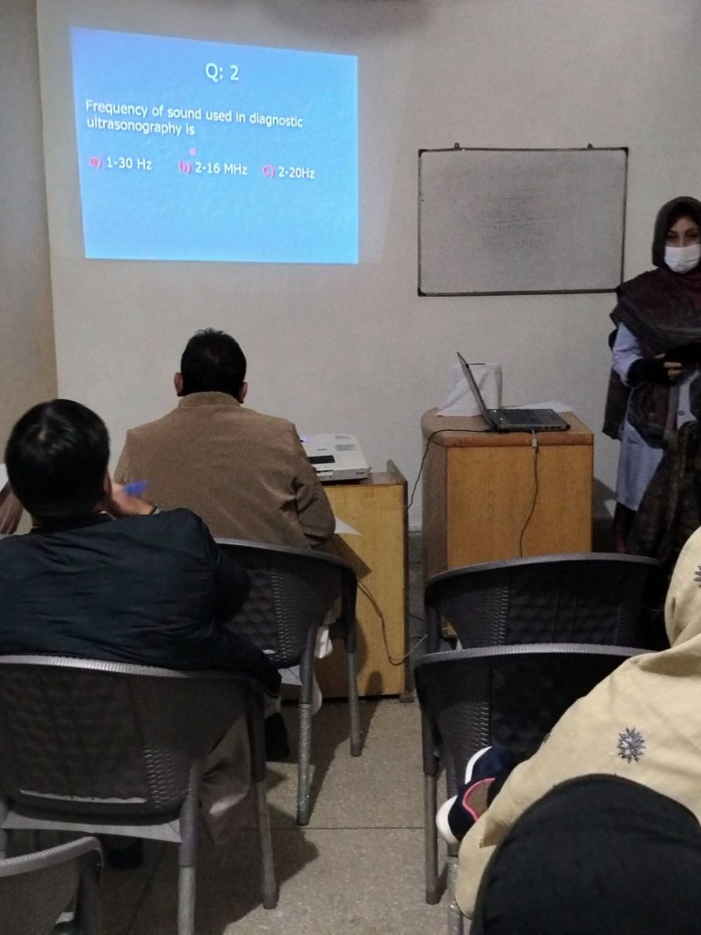
Hands-On Practical Training
We emphasize real-time scanning practice on patients and simulation models, under the direct supervision of experienced faculty.
Each participant will have dedicated time for live scanning, interpretation, and case discussions to build both skill and confidence.
Enroll Now
Join a growing community of healthcare professionals advancing their practice through ultrasound.
Limited Seats Available.
Duration: 06 Weeks (30 Working Days)
Visiting Fellowship in Diagnostic Ultrasound
Advance your diagnostic imaging skills and deepen your clinical expertise through a comprehensive and immersive Visiting Fellowship in Diagnostic Ultrasound. Tailored for medical professionals seeking to enhance their practical knowledge and experience in ultrasound diagnostics, this program offers a unique opportunity to engage with real patients, observe a wide spectrum of pathologies, and perform ultrasound examinations under the guidance of seasoned ultrasound specialists.
Program Highlights
Flexible Duration:
Choose a fellowship length that suits your availability and learning goals — whether you’re looking for an intensive one-month exposure or an in-depth, year-long clinical immersion.
Extensive Clinical Exposure:
Participants will gain firsthand experience by observing and assisting with ultrasound examinations on:
• Normal patients to understand baseline anatomy and scanning protocols
• Patients with common pathologies (e.g., gallstones, liver disease, kidney cysts, thyroid nodules)
• Patients with rare and complex pathologies, providing valuable insight into uncommon clinical presentations
Comprehensive Areas of Focus:
The fellowship encompasses a wide range of diagnostic ultrasound specialties, including:
• Abdominal ultrasound: liver, gallbladder, kidneys, pancreas, spleen
• Cranial ultrasound: particularly useful in neonates and infants
• Small parts ultrasound: thyroid, scrotum, breast, salivary glands
• Vascular ultrasound: including screening and evaluation for:
• Deep Vein Thrombosis (DVT)
• Carotid artery disease
• Peripheral arterial and venous systems
Hands-On Training:
Beyond observation, fellows will be actively engaged in hands-on scanning, gaining practical experience in:
• Patient positioning and preparation
• Probe handling and real-time image acquisition
• Identifying normal and abnormal findings
• Case-based learning and image interpretation
Expert Supervision & Mentorship:
Work alongside highly experienced ultrasound instructors who will provide constant guidance, feedback, and mentoring throughout your fellowship.
Case Discussions & Learning Modules:
Benefit from structured teaching sessions, case reviews, and interactive discussions to solidify both your technical and clinical decision-making skills.
Who Should Apply?
This fellowship is ideal for:
• Medical doctors (MBBS or MD) seeking additional clinical experience in ultrasound
• Radiology residents or general practitioners aiming to expand their diagnostic capabilities
• International medical graduates interested in comparative clinical practice
Certification:
A certificate of completion will be awarded based on attendance, performance, and duration of training.
Apply Now
Spaces are limited to ensure personalized instruction.
Start your journey toward advanced diagnostic proficiency today.
Duration: Flexible — from 1 Month up to 1 Year
Certificate in Diagnostic Ultrasound
The Six-Month Certificate in Diagnostic Ultrasound is a comprehensive training program designed for medical professionals who wish to gain in-depth theoretical knowledge and practical skills in the field of diagnostic sonography. This structured course integrates didactic learning, hands-on scanning, and clinical interpretation — equipping participants with the competence and confidence to independently perform and interpret a wide range of ultrasound examinations.
Program Highlights
Eligibility Criteria
This program is intended for:
• Medical Doctors (MBBS or MD)
• House Officers, Residents, or Practicing Physicians in specialties such as Radiology, Internal Medicine, Obstetrics & Gynecology, Surgery, or General Practice
• International Medical Graduates with equivalent qualifications
Note: Prior experience in ultrasound is not required, but basic knowledge of human anatomy and clinical medicine is essential.
Course Structure & Curriculum
The curriculum is divided into theory modules, practical demonstrations, and supervised clinical practice, focusing on the following key areas:
Ultrasound Physics & Instrumentation
• Basics of sound waves and image formation
• Machine settings, probe selection, and scanning techniques
• Understanding artifacts and optimizing image quality
Abdominal Ultrasound
• Liver, gallbladder, pancreas, spleen, kidneys, urinary bladder
• Aorta and major abdominal vessels
• Common pathologies (e.g., stones, masses, cysts)
Pelvic & Obstetric Ultrasound
• Non-pregnant and pregnant uterus
• Ovarian and adnexal assessments
• First-trimester pregnancy and fetal viability
• Basic fetal biometry and anomaly screening
Small Parts Ultrasound
• Thyroid and parathyroid glands
• Breast, scrotum, and superficial lumps
• Musculoskeletal basics
Vascular Ultrasound
• Evaluation for Deep Vein Thrombosis (DVT)
• Carotid Doppler and plaque assessment
• Systematic reporting formats
• Correlating clinical data with imaging findings
• Error recognition and differential diagnosis
Practical Hands-On Training
Hands-on training is a core component of the course and includes:
• Supervised patient scanning in hospital and outpatient settings
• Real-time imaging on both normal volunteers and patients with known pathologies
• Skill stations and simulated cases for learning probe control and anatomy localization
• Case-based discussions and weekly image review sessions
• One-on-one mentorship from expert radiologists and senior sonographers
Participants will gradually move from assisted scanning to independent image acquisition and preliminary reporting under supervision.
Assessment & Certification
• Mid-course and Final Assessment: Includes written tests, viva, and practical scanning evaluations
• Attendance Requirement: Minimum 80% attendance required
• On successful completion, participants will receive a Certificate in Diagnostic Ultrasound from Hameed Gul Memorial Ultrasound Institute
Why Choose This Course?
• Structured curriculum tailored to practical clinical application
• Extensive hands-on exposure with real patients
• Faculty with years of diagnostic imaging experience
• Supportive, academic environment with continuous feedback
• Strong foundation for those planning careers in radiology or general ultrasound practice
Duration: 06 Months
Focused Echocardiography in Emergency Life Support (FEEL)
Used to assess cardiac function in patients with cardiac arrest. This coarse is designed to provide you with a focus on sonographic planes, terminology and probe manipulation skills, along with techniques involved in handheld ultrasound Echocardiography to evaluate the heart chambers in 2D. Learn how to differentiate normal heart function from major cardiovascular pathology in this focus tutorial. The professional will also identify situations when a referral for a second opinion is indicated.
Duration: one Day
Bedside Lung Ultrasound in Emergency (BLUE)
Used to assess for fluid in the lungs. Lung. In a first glance Prof. Dr Nasir Hameed will talk about pleural effusions. Next to come are artifacts of the lung such as A-lines and B-lines, how to identify a pulmonary edema and you will learn how you can quickly and accurately identify a Pneumothorax.
Duration: one Day
Point-of-care ultrasound (POCUS)
POCUS is the use of ultrasound imaging at the bedside or point of care, allowing clinicians to perform and interpret ultrasound scans immediately. POCUS aims to improve diagnostic accuracy and expedite patient management by enabling quick bedside assessments.
Program Highlights…
Key features of POCUS:
Bedside/point of care:
POCUS is performed at the patient’s location, whether it’s a hospital bed, ambulance, or even a remote village.
Clinician-performed:
The ultrasound is typically performed by a trained clinician (e.g., physician, nurse, etc.) rather than a radiologist.
Immediate clinical integration:
The ultrasound findings are immediately used to guide clinical decisions and patient care.
Portability:
POCUS systems are often portable, allowing them to be easily moved to the patient’s location.
Enhanced diagnostics:
POCUS can complement physical examination and improve diagnostic accuracy, particularly in emergency and critical care settings.
Improved efficiency:
POCUS can reduce the need for other imaging modalities, such as CT scans, and shorten diagnostic delays.
Benefits of POCUS:
Faster diagnosis:
POCUS allows for quicker identification of conditions and faster initiation of treatment.
Improved patient outcomes:
By enabling earlier and more accurate diagnosis, POCUS can lead to better patient outcomes.
Scanning the lungs is not difficult to learn and it can easily be combined with the ultrasound of other organs. This course covers the sonographic anatomy and demonstrates how to image the lungs. We provide you with in-depth insights into pleural effusion, pulmonary congestion, consolidations and pneumothorax
Duration: One Week
Musculoskeletal
Point-of-care ultrasound can also be used in assessing musculoskeletal injuries and disorders. It’s especially useful for evaluating soft tissues like tendons and ligaments, and can guide joint injections or aspirations. This course provides an introduction to the anatomy and ultrasound examination of the knee and shoulder. Particular emphasis is given to scanning protocols as well as tips and strategies for obtaining high-quality images. The professional will be able to identify the sonographic appearance of normal and abnormal structures required to answer common clinical questions and understand situations when a referral for a second opinion is indicated.
Duration: One Day
Focused Assessment with Sonography for Trauma (FAST)
Used in trauma patients to assess for free fluid in the abdomen. Here we teach you the FAST protocol, which is used in the ultrasound assessment of trauma patients. We will explain when it should be used and which views you should acquire. Learn which pathologies you can detect and watch teaching cases, which highlight the potential of the FAST exam.
Duration: One Day
Assessment for DVT
This course aims to improve your understanding and use of handheld ultrasound to answer clinical questions by focussing on sonographic planes, terminology, and probe manipulation skills. Importantly, an overview of anatomical landmarks of the peripheral venous system is included as part of this course. The professional will recognise the variants and abnormalities of the peripheral venous system from the presentation of a hands-on demo and case-based learning and will understand situations when a referral for a second opinion is indicated.
Duration: Two Days
Carotid Doppler
Imaging the carotid arteries is “straight forward’. In this course we will explain, which instrumentation you need and how a carotid ultrasound exam is performed. Here you will also review the anatomy of neck arteries and learn when a carotid scan is indicated in a point of care setting. After completion you will know how to assess the Intima Media Thickness (IMD) and detect plaques to assess cardiovascular risk. In addition, we cover the topic of “carotid artery dissection”.
Duration: Two Days
Star Students / Alumni Success Stories
Recognizing excellence and outstanding achievements among our students. These students have demonstrated exceptional skills and dedication to the fields of ultrasound diagnostics.
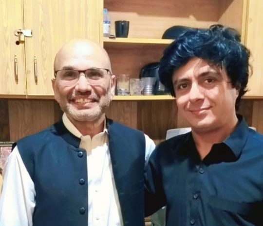
Dr. Sadiq Hussain
Dr. Hussain Ali
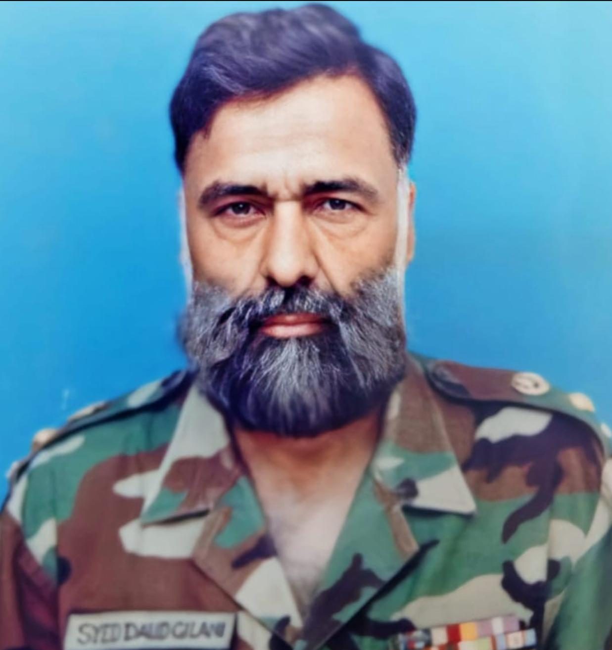
Colonel (Rtd)
Dr. Syed Daud Shah Gilani
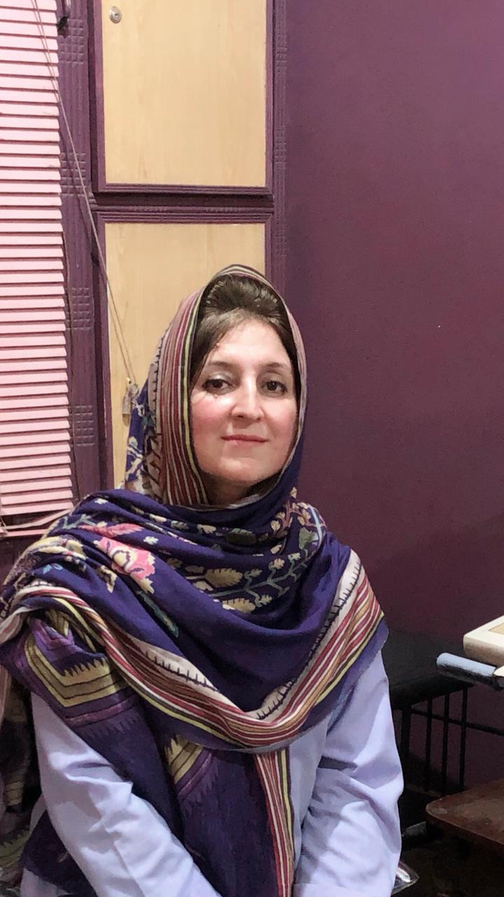
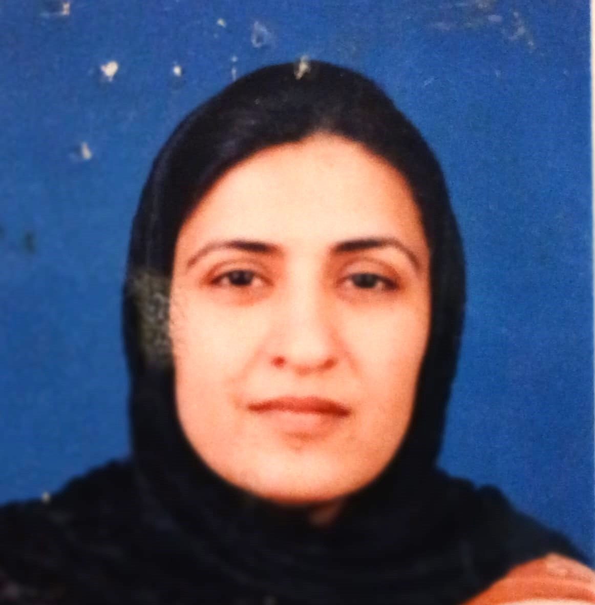
Dr Yasmin Khattak
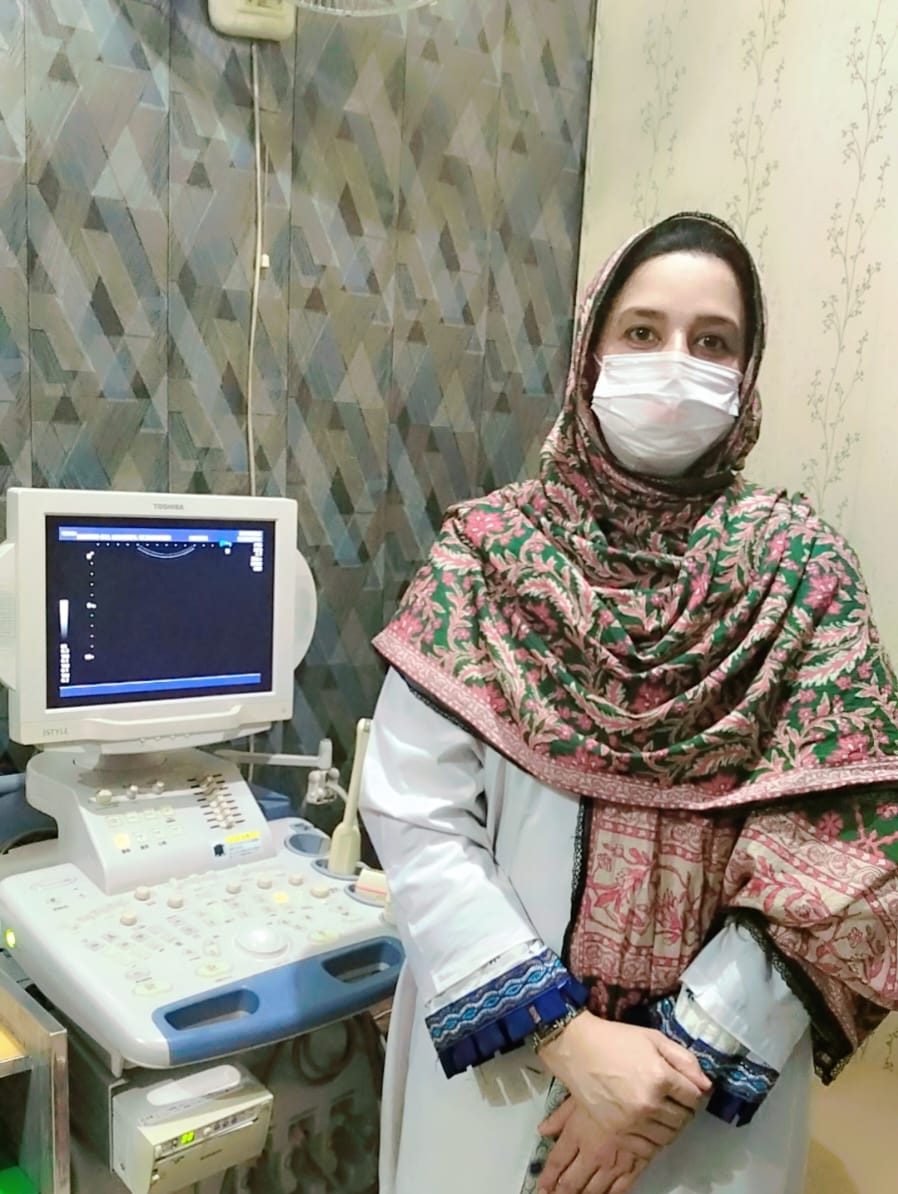
Dr Muzamila Salim
Mrs. Zelikha
Our Alumni Network
Join the global community of ultrasound professionals who have graduated from our instittute and are making significant contributions to healthcare.
Alumni Success Stories
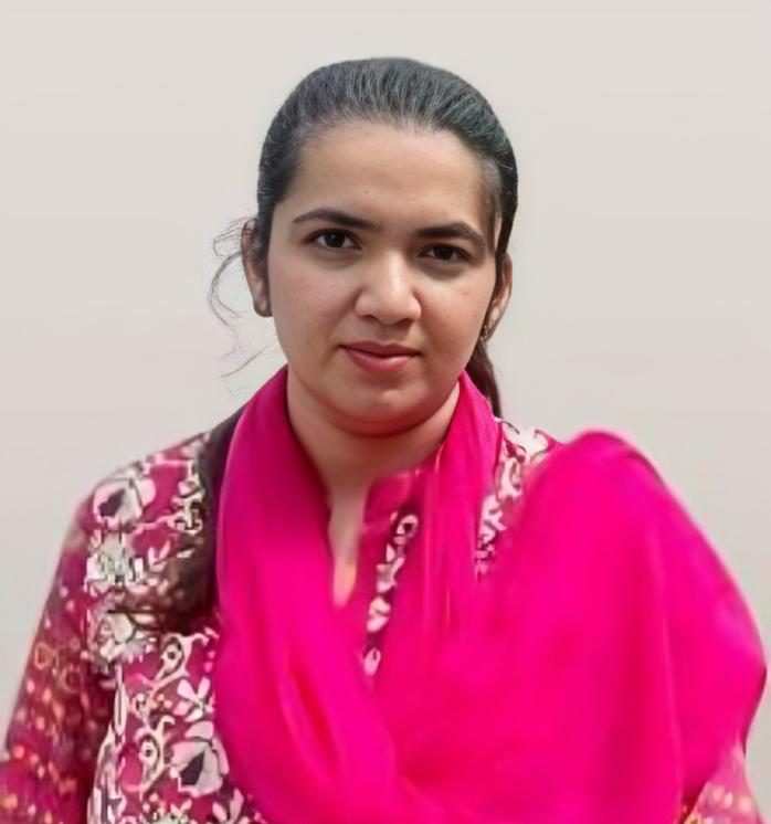
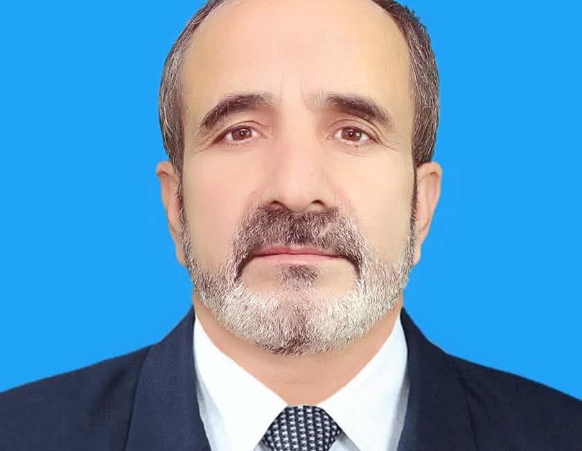
Dr. Saeeda Gul
(Afternoon HGMUIC DIR Medical Centre Branch)